需要订阅 JoVE 才能查看此. 登录或开始免费试用。
Method Article
经支气管肺冷冻活检诊断间质性肺疾病和外周肺病变 - 一种阶梯式方法
摘要
经支气管肺冷冻活检 (TBLC) 用于诊断间质性肺疾病和外周肺病变是一种高产诊断和安全的手术。我们描述了一种使用可弯曲支气管镜针对提到的不同适应症进行 TBLC 的逐步方法,这可能对进行 TBLC 的新手支气管镜医生有所帮助。
摘要
经支气管肺冷冻活检 (TBLC) 是一种侵入性手术,在过去十年中越来越多地实施,作为电视辅助胸外科肺活检 (SLB) 的替代方法,用于诊断间质性肺疾病 (ILD)。TBLC 的适应症主要是在根据先前的多学科团队讨论无法实现时对特定 ILD 亚型进行亚分类。尽管 SLB 被认为是建立组织学诊断的金标准,但由于 TBLC 的诊断率与 SLB 相当,但就并发症(包括死亡率)而言,TBLC 已逐渐被建议作为未分类 ILD 患者的首选组织学诊断方式。近年来,桡动脉支气管内超声 (R-EBUS) 和电磁导航支气管镜 (ENB) 引导下治疗外周肺病变的 TBLC 也被描述为安全的手术,与镊子活检相比,它可能提高诊断率。尽管如此,TBLC 的诊断特性仍取决于手术性能的质量。本文旨在描述使用柔性支气管镜针对上述不同适应症进行 TBLC 的逐步方法,这可能对进行 TBLC 的新手支气管镜医生有所帮助。
引言
间质性肺病 (ILD) 由一组急性和慢性肺部疾病组成,影响形成间质的所有肺实质成分中的一个或多个,例如支气管、肺泡、结缔组织以及血液和淋巴管。尽管是罕见疾病,但 ILD 的 200 多种不同亚型代表了具有不同临床、放射学和细胞组织学特征的异质性疾病类别。ILD 通常表现为炎症、纤维化或两者兼而有之,这是患者通常感知症状(如干咳、劳力性呼吸困难和疲劳)的根本原因 1,2。
ILD 分为特发性间质性肺炎 (IIP)、已知病因的间质性肺炎(例如结缔组织病间质性肺病、药物诱导的 ILD 和工作相关性尘肺病)、肉芽肿性间质性肺炎(例如结节病和过敏性肺炎)和孤儿 ILD(例如,多发性囊性肺病和嗜酸性粒细胞性肺炎)1.这种分类和进一步的诊断亚型对于确定最佳治疗和随访以及预后预测至关重要。然而,由于诊断难题可能具有挑战性,建议对基于多学科团队讨论 (MDD) 获得的可用临床(包括病史、处置和潜在暴露)和辅助临床信息进行解释,如胸部高分辨率计算机断层扫描 (HRCT)、肺生理学和自身免疫学 3,4,5。如果无法获得可靠的 MDD 诊断 6,7,则使用经支气管肺冷冻活检 (TBLC) 进行组织学取样以增加明确 ILD 亚型诊断的可能性 8,9。在精心挑选的患者中,TBLC 被认为是一种安全的侵入性手术,其诊断准确性接近电视辅助胸外科肺活检 (SLB),后者仍被视为组织学 ILD 诊断的组织学金标准 10,11,12,13,14.TBLC 程序作为系统支气管镜检查进行,应用特殊的冷冻探针进行组织学采样,并在推荐的透视引导下进行。建议在使用 MDD 设置的三级 ILD 中心进行 TBLC,并由熟悉 TBLC 并发症管理的介入肺科医生进行,他们已在具有 TBLC 专业知识的专门中心接受了培训 9,10,11,15,16,17。
TBLC 最近也作为一种与桡支气管内超声 (R-EBUS) 相结合用于 ILD 诊断的程序而受到关注18,19。此外,与传统的经支气管钳活检相比,TBLC 已与 R-EBUS 和电磁导航支气管镜检查 (ENB) 联合用于诊断外周肺病变 (PPL),以提高诊断率20,21。然而,这种相对较新颖的 PPL 诊断方法尚未作为标准程序实施,因此需要该特定领域的进一步证据。本报告的目的是描述在临床环境中使用可弯曲支气管镜针对上述适应症进行 TBLC 的逐步方法。
研究方案
作者来自丹麦的两个 TBLC 中心(欧登塞大学医院和奥胡斯大学医院),这两个中心都根据赫尔辛基宣言的原则进行研究。伦理批准不是必需的,因为该研究本质上是观察性的。所有用于研究目的的患者都给出了书面知情同意书。需要强调的是,所描述的 TBLC 电导率的逐步方法与软性支气管镜的使用有关,并且基于国际指南的建议、专家声明、最先进的评论以及两个 TBLC 中心的经验 9、10、11、15、16、17、22、23、24,25.
1. PreTBLC 注意事项
- 确保有 TBLC 的指征,这对于在涉及肺科医生和放射科医生的先前 MDD 中整合来自 HRCT、生物化学和自身免疫学的信息无法建立可信的 ILD 诊断的患者来说是合理的。
- 通过避免 表 1 中描述的禁忌证来选择合适的患者。
| 相对禁忌证 | 绝对禁忌证 |
| 用力肺活量 (FVC) <预测值的 50% | 血小板减少症 < 50 x 109/L 或 INR > 1.5 |
| 肺一氧化碳扩散量 (DLCO) <预测值的 35% | 未矫正的出血素质 |
| 收缩期肺动脉压 > 50 mmHg(例如,根据超声心动图估计) | 由于肺功能受损患者并发症风险增加而导致的进行性和临床下降 |
| 体重指数 > 35 kg/m2 |
表 1:TBLC 的禁忌症。 TBLC 电导的相对和绝对禁忌症。缩写:TBLC = 经支气管肺冷冻活检。
2. PreTBLC 制备
- 回顾 HRCT 和胸部放射科医生的建议,以根据放射学疾病表现规划最容易获得哪些支气管节段 (BS) 组织学取样。
- 在 TBLC 性能之前测试系统是否正常工作。
- 按下设置面板上的气体(二氧化碳 (CO2) 或一氧化二氮 (NO))油箱容量按钮,检查气瓶中的气体体积。
- 将低温探头放在托盘上,在按下踏板脚开关 5-10 秒的同时观察探头。在探头尖端寻找一个冰球,表明它运行正常(图 1)。
- 在 TBLC 下使用全身麻醉 (GA) 或深度镇静,并考虑用 0.5-1 g 氨甲环酸进行术前用药,以降低出血风险。
- 在气管中放置一根 7.5-8.5 毫米尺寸的特殊双腔气管插管 (ETT)。
注意:ETT 有一个主通道,允许在患者通气时进入支气管镜,还有一个次要侧通道,用作支气管阻滞剂导管的工作通道。- 持续喷洒局部麻醉剂(例如,10% 利多卡因喷雾剂)以减轻咳嗽。另请参见步骤 3.5。
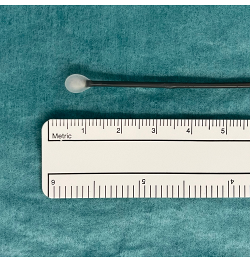
图 1:冰球表示可用的 TBLC 设备。 踏板激活 CO2 气体从水箱中扩散并诱导冻结。这是在水中测试的,如果功能正常,冰球会出现在低温探针的尖端。缩写:TBLC = 经支气管肺冷冻活检。 请单击此处查看此图的较大版本。
3. TBLC 电导率
- 通过 ETT 引入可弯曲支气管镜并执行支气管镜检查程序。
注意:一些 TBLC 中心使用硬质支气管镜来插管气管。如果气管由刚性支气管镜插管,则可以将柔性支气管镜穿过刚性支气管镜。- 将柔性冷冻探针通过支气管镜的工作通道引入选定的 BS 中。
注:冷冻探针既有一次性探针(1.1、1.7 和 2.4 mm)也有可重复使用的探针(1.9 和 2.4 mm)。 - 使用透视确保冷冻探针尖端的位置距离对应于所选 BS 的胸壁约 10 毫米(图 2)。
- 在双腔 ETT 的侧通道中引入支气管阻滞导管(例如 Fogarty 球囊),并将其放置在选定的 BS 口(图 3)。
- 给支气管阻滞剂导管充气,以评估相对于球囊远端发生的潜在出血的放置和阻塞的适当性(图4)。
- 如果球囊放置正确,请给支气管阻滞剂导管放气。
- 用 pean 固定支气管阻滞剂导管,确保球囊的位置。
- 在相应的 ETT 及其侧通道中使用少量利多卡因喷雾剂或生理盐水,以减少因在 ETT 中引入冷冻探针和 ETT 侧通道中引入球囊导管而产生的任何摩擦。
- 当步骤 3.1.1-3.1.3 令人满意地执行时,根据冷冻探针的大小,踩下冷冻踏板 3-6 秒,以利用焦耳-汤普森定律将肺实质组织冷冻至约 -45-79 °C(对于 CO2 )和 -89 °C(对于 NO)。
- 快速缩回包含冷冻探头的柔性支气管镜,同时按住冷冻踏板以继续冷冻并防止活检在回缩过程中脱落。
- 在步骤 3.1.5 中描述的作期间,让支气管镜医师以外的人保持球囊充气,以堵塞所选 BS 口远端的活检部位,以控制潜在的出血。
- 将柔性冷冻探针通过支气管镜的工作通道引入选定的 BS 中。
- 继续步骤 3.1.6,直到从同一肺叶的两个 BS 中获得至少两次活检。
- 将活检结果放入盐水中,获得所有活检结果后,将它们固定在甲醛 (4%) 中(图 5)。
- 在 MDD 之前将活检送去进行病理检查。
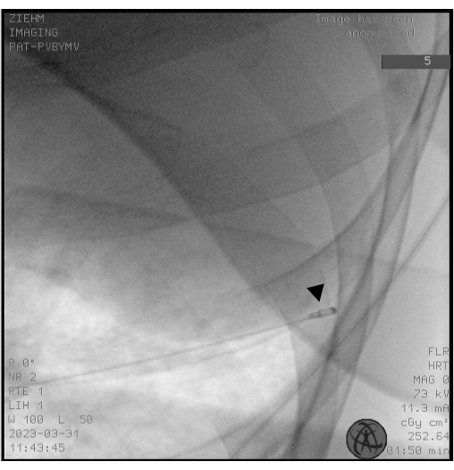
图 2:透视。透视用于确保在冷冻前正确放置冷冻探针。 低温探针的尖端显示为鼓槌的头部(黑色箭头)。 请单击此处查看此图的较大版本。
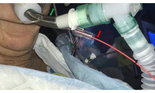
图 3:气管插管。 双腔气管插管(绿色箭头)允许支气管镜进入气道,同时通过在侧通道中引入球囊导管(红色箭头)来控制出血。 请单击此处查看此图的较大版本。

图 4:球囊导管的充气。 在进行经支气管肺冷冻活检后,球囊导管充气以确保阻塞并防止球囊远端分布到肺叶其他部分的潜在出血。 请单击此处查看此图的较大版本。
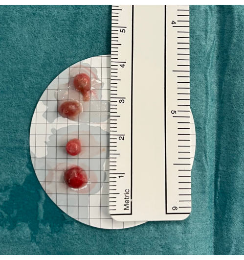
图 5:活检。 经支气管肺冷冻活检置于冷盐水中,然后固定在甲醛中。 请单击此处查看此图的较大版本。
4. TBLC 后程序
- 每次活检后,将支气管镜重新引入 BS 并给球囊放气,观察是否出血。
- 如果观察到出血,请重新充气球囊(图6)。如果球囊处于正确位置并阻塞了 BS,请等待几分钟让出血停止,然后继续 TBLC 程序。
- 如果在步骤 4.1.1. 后仍观察到出血,请在球囊远端安装冰冷的盐水。
- 如果步骤 4.1.2 后持续出血。或血液溢出到其他 BS 中的球囊衰竭,结合使用抽吸、支气管内注射冰冷的生理盐水(含或不含肾上腺素)和氨甲环酸。
- 如果凝固的血液阻塞了气道,请使用冷冻探针再次打开气道,方法是将冷冻探针的尖端冻结到血凝块中,然后将其通过 ETT 向上缩回。
- 如果出血仍未得到控制,请将 ETT 更改为既能使未活检的肺通气又能阻塞出血肺上的主要支气管的 ETT,并将患者转移到重症监护病房。
- 在 TBLC 后进行聚焦肺部超声 (FLUS),同时患者仍处于镇静状态,以确定医源性气胸 (PTX) 的指征。
- 如果 FLUS 观察表明 PTX 的可能性很高,则考虑使用尾纤导管 (Fr 7-16) 插入 FLUS 引导下的胸膜引流管。如果 FLUS 提示 PTX 大小迅速增加,尤其是患者的临床状况恶化,则需要胸腔引流。
- 如果 FLUS 显示小 PTX 且患者临床稳定,则推迟拔管 5-10 分钟。如果 PTX 大小此后进展或患者临床不稳定,请在拔管前插入胸膜引流管。
- 如果没有 PTX 迹象或 PTX 大小进展并且患者临床稳定,请在步骤 4.2 或步骤 4.2.2 后为患者拔管。
- 在 TBLC 后在恢复室观察患者。在临床情况稳定的患者回家之前,确保重复 FLUS 或胸部 X 线检查不存在迟发性 PTX。
- 与肺科医生、放射科医生和病理学家讨论后续 MDD 活检的组织学特征表现,并结合其他细节,以得出具有高诊断概率的 ILD 亚型。
- 在门诊告知患者第 4.5 步的 MDD 结论,并计划可能的治疗和随访。
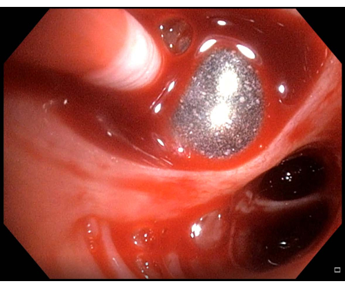
图 6:轻微出血。 如果在进行经支气管肺冷冻活检后观察到出血,在这种情况下,轻微出血,应在再次放气前几分钟保持球囊导管充气。 请单击此处查看此图的较大版本。
5. TBLC 与 R-EBUS 和 ENB 联合用于 PPL 诊断
- 导航并确认外周肺病变的位置。
- 在透视的引导下将 1.1 mm 冷冻探针插入扩展的工作通道中。
- 将凝固探头的尖端与径向 EBUS 探头的位置相匹配。
- 踩下冷冻踏板 4-8 秒。
- 通过扩展的工作通道快速缩回冷冻探针,同时按住冷冻踏板以继续冷冻并防止活检在回缩过程中脱落。
- 在缩回冷冻探针时,保持支气管镜和扩展工作通道就位。
- 重复步骤 5.2-5.4,直到获得足够数量的活检。
- 按照步骤 3.3 和 3.4 中的说明处理活检。
结果
根据作者来自两个 TBLC 中心的观察,所描述的带有柔性支气管镜的 TBLC 的逐步程序允许对精心选择的丹麦患者进行组织学取样,这些患者尽管之前有 MDD,但仍未被诊断为 ILD 亚型。最近发表的两项研究23,25 报告了这些队列的详细观察结果,第一作者的中心总结了表 2。
讨论
无论 TBLC 的适应症如何,其诊断特性都取决于手术性能的质量和接受手术的选择标准。这强调了实施正式和经认证的培训计划的建议,以获得执行标准化 TBLC 程序所需的能力。尽管目前无法获得官方的 TBLC 教育,但最近欧洲呼吸学会关于 ILD TBLC 的指南建议,进行 TBLC 的介入肺科医生应接受正式培训,以提高诊断率并降低并发症风险,同时还能够管理这些并发症,例如,?...
披露声明
作者没有需要披露的利益冲突。
致谢
作者要感谢丹麦欧登塞大学医院支气管镜病房胸外科和麻醉科的工作人员,感谢他们为本文准备图表提供的帮助。
材料
| Name | Company | Catalog Number | Comments |
| "Chimney" for tube | |||
| CO2 gas bottle adapter | |||
| CO2 gas tank | Erbe | ||
| Endoscopy column | |||
| Endotracheal tube, size 7.5-8.5 mm | Erbe | ||
| Erbecryo pedal footswitch | Erbe | ||
| Erbecryo2 workstation | Erbe | ||
| Flexible bronchoscope | |||
| Flexible gas hose | Mediland | ||
| Flexible single use cryoprobe, OD 1.1 mm | Erbe | ||
| Flexible single use cryoprobe, OD 1.7 mm | Erbe | ||
| Flexible single use cryoprobe, OD 2.4 mm | |||
| Fluoroscope | |||
| Fogarty balloon catheter | |||
| Formalin glasses in closed system | |||
| NaCl incl. cold NaCl | |||
| Pean for fixating Fogarty balloon | |||
| Sterile disposable cup | |||
| Sterile suction tube | |||
| Sterile tweesers | |||
| Syringe for Fogarty balloon inflation/deflation | |||
| Table bag for flouroscope | |||
| Three way tap for Fogarty balloon syringe | |||
| Tracheal suction | |||
| Ultrasound machine | Erbe | ||
| Valve for biopsy chanel | |||
| Valve to suction duct |
参考文献
- Travis, W. D., et al. An official American Thoracic Society/European Respiratory Society statement: Update of the international multidisciplinary classification of the idiopathic interstitial pneumonias. American Journal of Respiratory and Critical Care Medicine. 188 (6), 733-748 (2013).
- Ruaro, B., et al. Editorial: Pulmonary fibrosis: One manifestation, various diseases. Frontiers in Pharmacology. 13, 1027332 (2022).
- Lamas, D. J., et al. Delayed access and survival in idiopathic pulmonary fibrosis: a cohort study. American Journal of Respiratory and Critical Care Medicine. 184 (7), 842-847 (2011).
- Tomassetti, S., Piciucchi, S., Tantalocco, P., Dubini, A., Poletti, V. The multidisciplinary approach in the diagnosis of idiopathic pulmonary fibrosis: a patient case-based review. European Respiratory Review. 24 (135), 69-77 (2015).
- Walsh, S. L. F., et al. Multicentre evaluation of multidisciplinary team meeting agreement on diagnosis in diffuse parenchymal lung disease: a case-cohort study. Lancet Respiratory Medicine. 4 (7), 557-565 (2016).
- Ryerson, C. J., et al. A standardized diagnostic ontology for fibrotic interstitial lung disease. An International Working Group perspective. American Journal of Respiratory and Critical Care Medicine. 196 (10), 1249-1254 (2017).
- Cottin, V., et al. Integrating clinical probability into the diagnostic approach to idiopathic pulmonary fibrosis: An International Working Group perspective. American Journal of Respiratory and Critical Care Medicine. 206 (3), 247-259 (2022).
- Rodrigues, I., et al. Diagnostic yield and safety of transbronchial lung cryobiopsy and surgical lung biopsy in interstitial lung diseases: a systematic review and meta-analysis. European Respiratory Review. 31 (166), 210280 (2022).
- Korevaar, D. A., et al. European Respiratory Society guidelines on transbronchial lung cryobiopsy in the diagnosis of interstitial lung diseases. European Respiratory Journal. 60 (5), 2200425 (2022).
- Colella, S., Haentschel, M., Shah, P., Poletti, V., Hetzel, J. Transbronchial lung cryobiopsy in interstitial lung diseases: best practice. Respiration. 95 (6), 383-391 (2018).
- Hetzel, J., et al. Transbronchial cryobiopsies for the diagnosis of diffuse parenchymal lung diseases: expert statement from the Cryobiopsy Working Group on safety and utility and a call for standardization of the procedure. Respiration. 95 (3), 188-200 (2018).
- Ravaglia, C., Poletti, V. Transbronchial lung cryobiopsy for the diagnosis of interstitial lung diseases. Current Opinion in Pulmonary Medicine. 28 (1), 9-16 (2022).
- Troy, L. K., et al. Diagnostic accuracy of transbronchial lung cryobiopsy for interstitial lung disease diagnosis (COLDICE): a prospective, comparative study. Lancet Respiratory Medicine. 8 (2), 171-181 (2020).
- Ruaro, B., et al. Transbronchial lung cryobiopsy and pulmonary fibrosis: A never-ending story. Heliyon. 9 (4), e14768 (2023).
- Lentz, R. J., Argento, A. C., Colby, T. V., Rickman, O. B., Maldonado, F. Transbronchial cryobiopsy for diffuse parenchymal lung disease: a state-of-the-art review of procedural techniques, current evidence, and future challenges. Journal of Thoracis Disease. 9 (7), 2186-2203 (2017).
- Maldonado, F., et al. Transbronchial cryobiopsy for the diagnosis of interstitial lung diseases: CHEST Guideline and Expert Panel Report. Chest. 157 (4), 1030-1042 (2020).
- Avasarala, S. K., Wells, A. U., Colby, T. V., Maldonado, F. Transbronchial cryobiopsy in interstitial lung diseases: State-of-the-art review for the interventional pulmonologist. Journal of Bronchology Interventional Pulmonology. 28 (1), 81-92 (2021).
- Abdelghani, R., Thakore, S., Kaphle, U., Lasky, J. A., Kheir, F. Radial Endobronchial Ultrasound-guided Transbronchial Cryobiopsy. Journal of Bronchology Interventional Pulmonology. 26 (4), 245-249 (2019).
- Inomata, M., et al. Utility of radial endobronchial ultrasonography combined with transbronchial lung cryobiopsy in patients with diffuse parenchymal lung diseases: a multicentre prospective study. BMJ Open Respiratory Research. 8 (1), e000826 (2021).
- Benn, B. S., Gmehlin, C. G., Kurman, J. S., Doan, J. Does transbronchial lung cryobiopsy improve diagnostic yield of digital tomosynthesis-assisted electromagnetic navigation guided bronchoscopic biopsy of pulmonary nodules? A pilot study. Respiratory Medicine. 202, 106966 (2022).
- Ankudavicius, V., Miliauskas, S., Poskiene, L., Vajauskas, D., Zemaitis, M. Diagnostic yield of transbronchial cryobiopsy guided by radial endobronchial ultrasound and fluoroscopy in the radiologically suspected lung cancer: A single institution prospective study. Cancers. 14 (6), 1563 (2022).
- Ravaglia, C., et al. Transbronchial lung cryobiopsy in diffuse parenchymal lung disease: Comparison between biopsy from 1 segment and biopsy from 2 segments - diagnostic yield and complications. Respiration. 93 (4), 285-292 (2017).
- Davidsen, J. R., Skov, I. R., Louw, I. G., Laursen, C. B. Implementation of transbronchial lung cryobiopsy in a tertiary referral center for interstitial lung diseases: a cohort study on diagnostic yield, complications, and learning curves. BMC Pulmonary Medicine. 21 (1), 67 (2021).
- Laursen, C. B., et al. Lung ultrasound assessment for pneumothorax following transbronchial lung cryobiopsy. ERJ Open Research. 7 (3), 00045-2021 (2021).
- Kronborg-White, S., et al. Integration of cryobiopsies for interstitial lung disease diagnosis is a valid and safe diagnostic strategy-experiences based on 250 biopsy procedures. Journal of Thoracic Disease. 13 (3), 1455-1465 (2021).
- Barisione, E., et al. Competence in transbronchial cryobiopsy. Panminerva Medica. 61 (3), 290-297 (2019).
- Raghu, G., et al. Idiopathic pulmonary fibrosis (an update) and progressive pulmonary fibrosis in adults: An Official ATS/ERS/JRS/ALAT Clinical Practice Guideline. American Journal of Respiratory and Critical Care Medicine. 205 (9), e18-e47 (2022).
- Ravaglia, C., et al. Diagnostic yield and risk/benefit analysis of trans-bronchial lung cryobiopsy in diffuse parenchymal lung diseases: a large cohort of 699 patients. BMC Pulmonary Medicine. 19 (1), 16 (2019).
- Hernandez-Gonzalez, F., et al. Cryobiopsy in the diagnosis of diffuse interstitial lung disease: yield and cost-effectiveness analysis. Archivos de Bronconeumología. 51 (6), 261-267 (2015).
- Cooley, J., et al. Safety of performing transbronchial lung cryobiopsy on hospitalized patients with interstitial lung disease. Respiratory Medicine. 140, 71-76 (2018).
- Hetzel, J., et al. Transbronchial cryobiopsy increases diagnostic confidence in interstitial lung disease: a prospective multicenter trial. European Respiratory Journal. 56 (6), 1901520 (2020).
- Kheir, F., et al. Transbronchial lung cryobiopsy in patients with interstitial lung disease: a systematic review. Annals of the American Thoracic Society. 19 (7), 1193-1202 (2022).
- Walscher, J., et al. Transbronchial cryobiopsies for diagnosing interstitial lung disease: real-life experience from a tertiary referral center for interstitial lung disease. Respiration. 97 (4), 348-354 (2019).
- Gnass, M., et al. Transbronchial lung cryobiopsy guided by radial mini-probe endobronchial ultrasound in interstitial lung diseases - a multicenter prospective study. Advances in Respiratory Medicine. 88 (2), 123-128 (2020).
- Ma, X., et al. Global and regional burden of interstitial lung disease and pulmonary sarcoidosis from 1990 to 2019: results from the Global Burden of Disease study 2019. Thorax. 77 (6), 596-605 (2022).
- Kronborg-White, S., et al. A pilot study on the use of the super dimension navigation system for optimal cryobiopsy location in interstitial lung disease diagnostics. Pulmonology. 29 (2), 119-123 (2021).
- Wijmans, L., et al. Confocal laser endomicroscopy as a guidance tool for transbronchial lung cryobiopsies in interstitial lung disorder. Respiration. 97 (3), 259-263 (2019).
- Kheir, F., et al. Using bronchoscopic lung cryobiopsy and a genomic classifier in the multidisciplinary diagnosis of diffuse interstitial lung diseases. Chest. 158 (5), 2015-2025 (2020).
- Renzoni, E. A., Poletti, V., Mackintosh, J. A. Disease pathology in fibrotic interstitial lung disease: is it all about usual interstitial pneumonia. Lancet. 398 (10309), 1437-1449 (2021).
- Chaudhary, S., et al. Interstitial lung disease progression after genomic usual interstitial pneumonia testing. European Respiratory Journal. 61 (4), 2201245 (2023).
- Raghu, G., et al. Use of a molecular classifier to identify usual interstitial pneumonia in conventional transbronchial lung biopsy samples: a prospective validation study. Lancet Respiratory Medicine. 7 (6), 487-496 (2019).
- Kheir, F., et al. Use of a genomic classifier in patients with interstitial lung disease: a systematic review and meta-analysis. Annals of American Thoracic Society. 19 (5), 827-832 (2022).
- Glenn, L. M., Troy, L. K., Corte, T. J. Novel diagnostic techniques in interstitial lung disease. Frontiers in Medicine. 10, 1174443 (2023).
- Kim, S. H., et al. The additive impact of transbronchial cryobiopsy using a 1.1-mm diameter cryoprobe on conventional biopsy for peripheral lung nodules. Cancer Research and Treatment. 55 (2), 506-512 (2023).
转载和许可
请求许可使用此 JoVE 文章的文本或图形
请求许可探索更多文章
This article has been published
Video Coming Soon
版权所属 © 2025 MyJoVE 公司版权所有,本公司不涉及任何医疗业务和医疗服务。